Pigmented BCC versus Pigmented Melanoma
Condition 1
Condition 2
Description
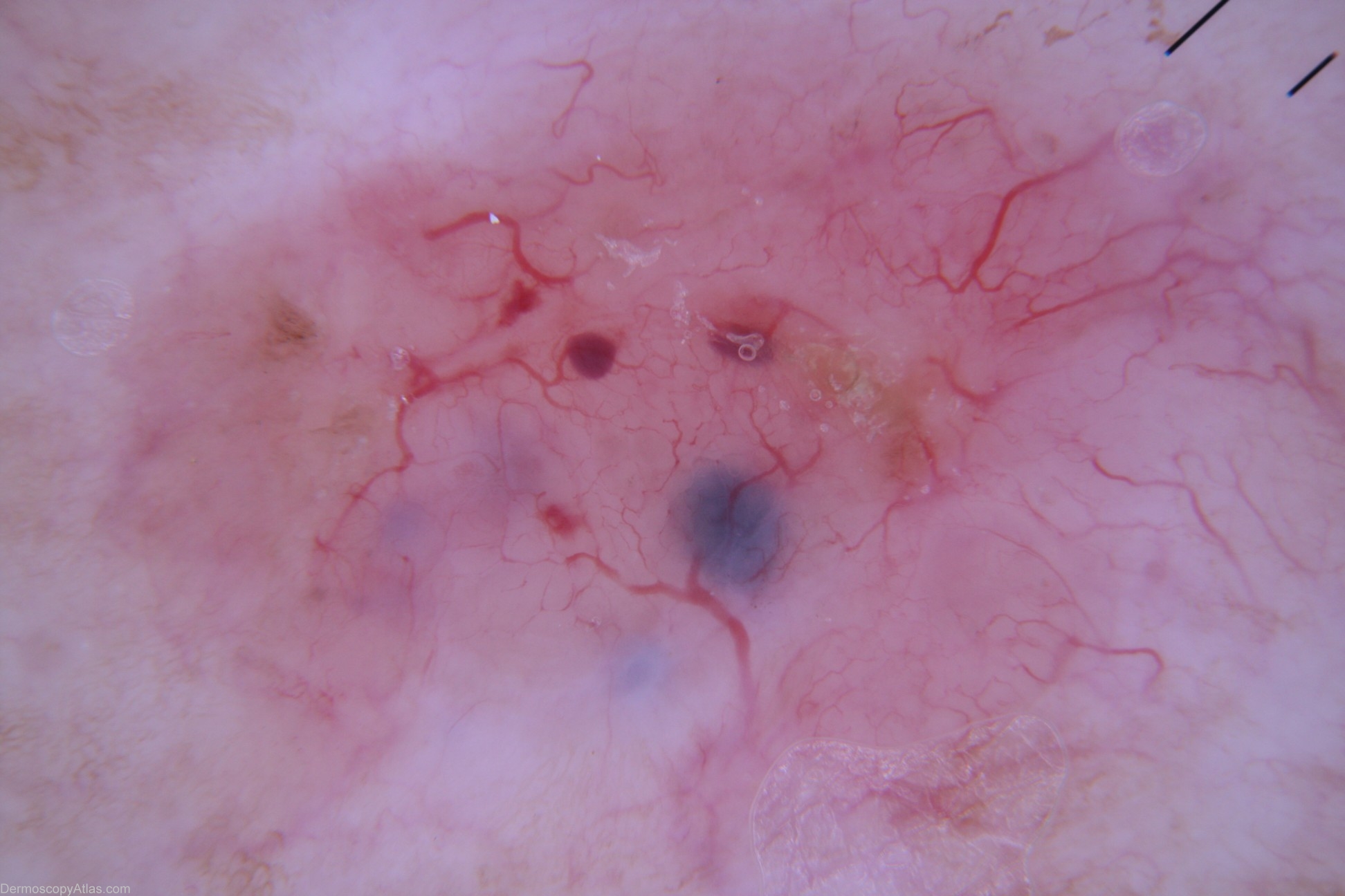
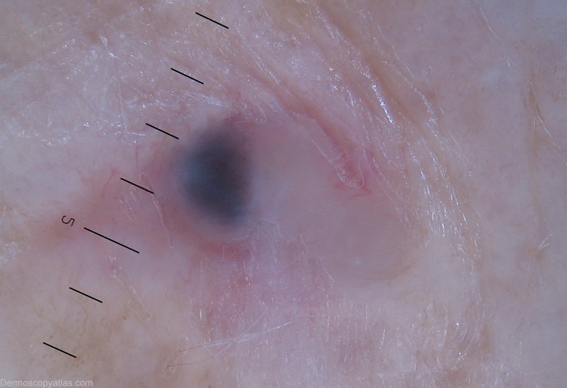
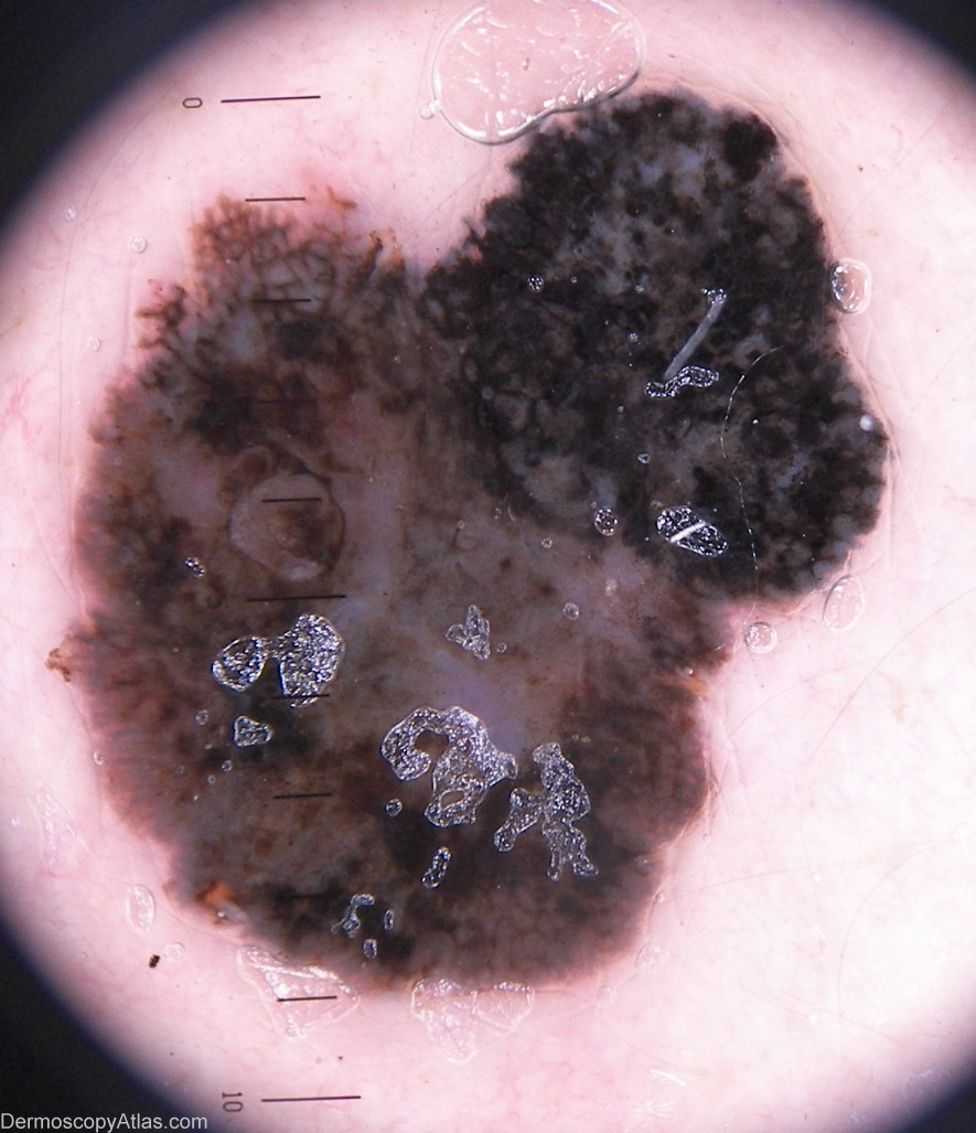
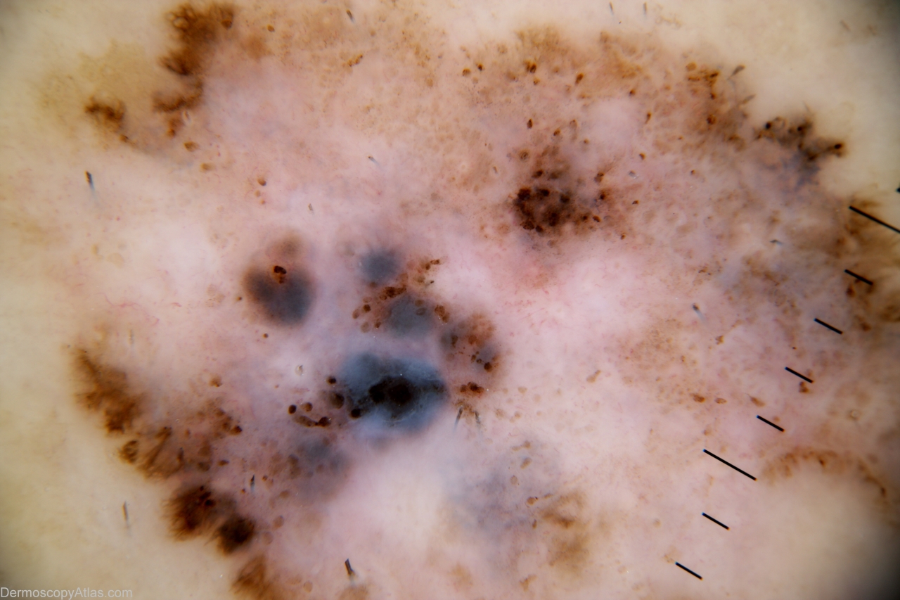
Pigmented Basal Cell Carcinoma (BCC) and melanoma are often indistinguishable to the naked eye, but dermoscopy reveals distinct architectural and vascular patterns that allow for reliable differentiation. The key to differentiating pigmented BCC from melanoma is the absence of a pigmented network and the presence of specific pigmented structures (clods dots and peripheral pigmented lines originating from a point) along with arborizing vessels.
Absence of a pigment network and arborising vessels are the best discriminators along with superficial ulceration to indicate a lesion is a BCC.
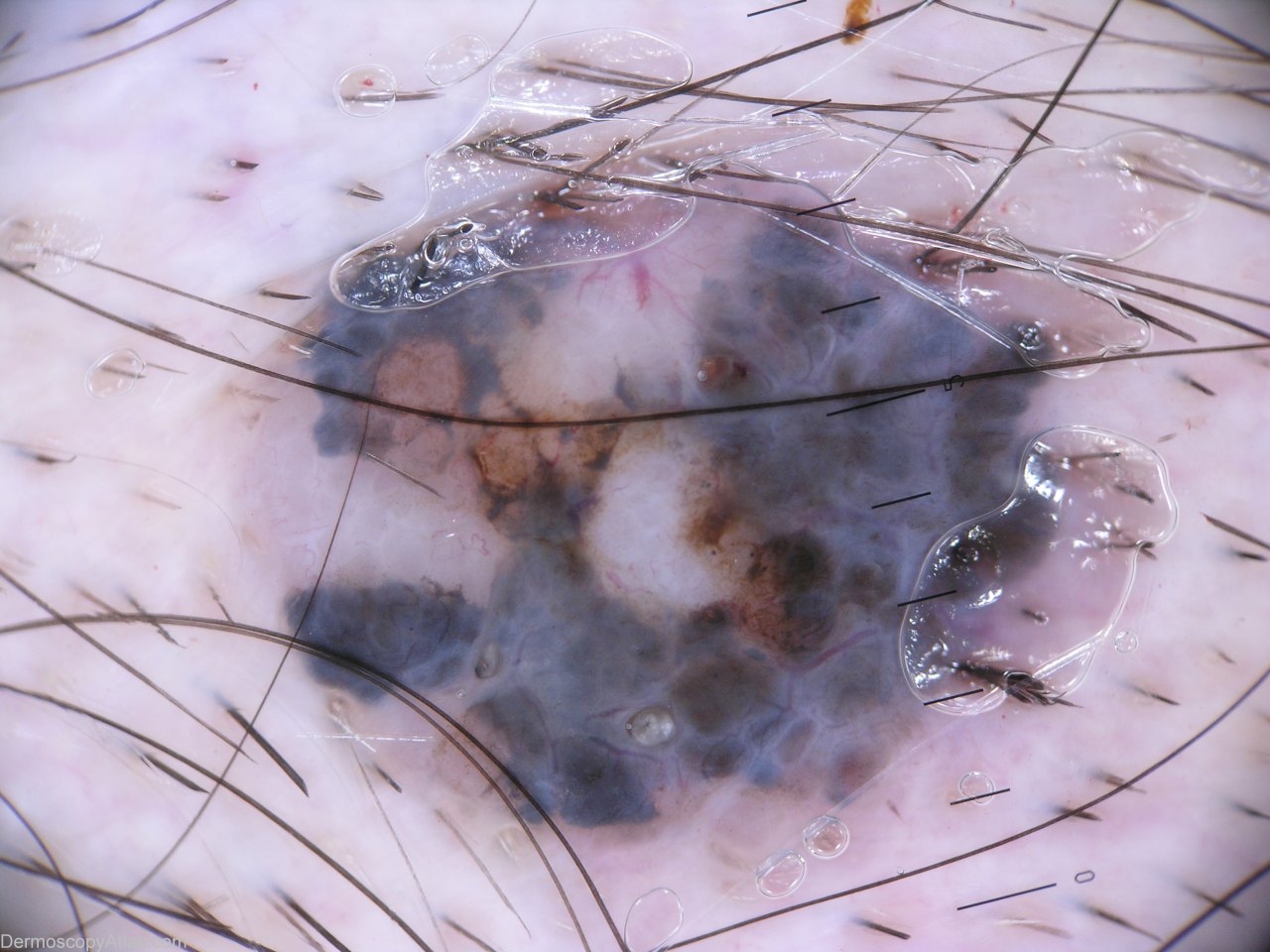
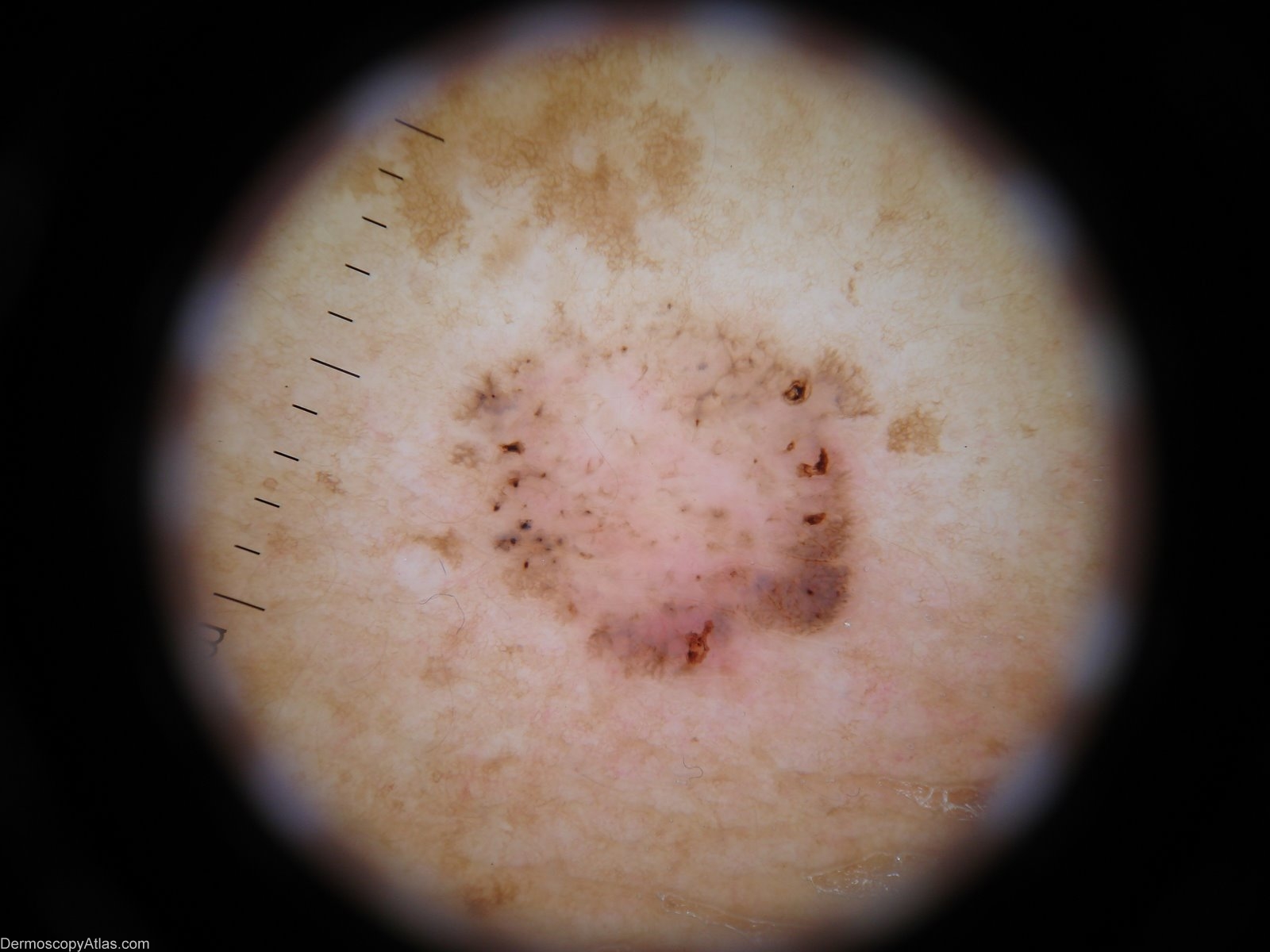
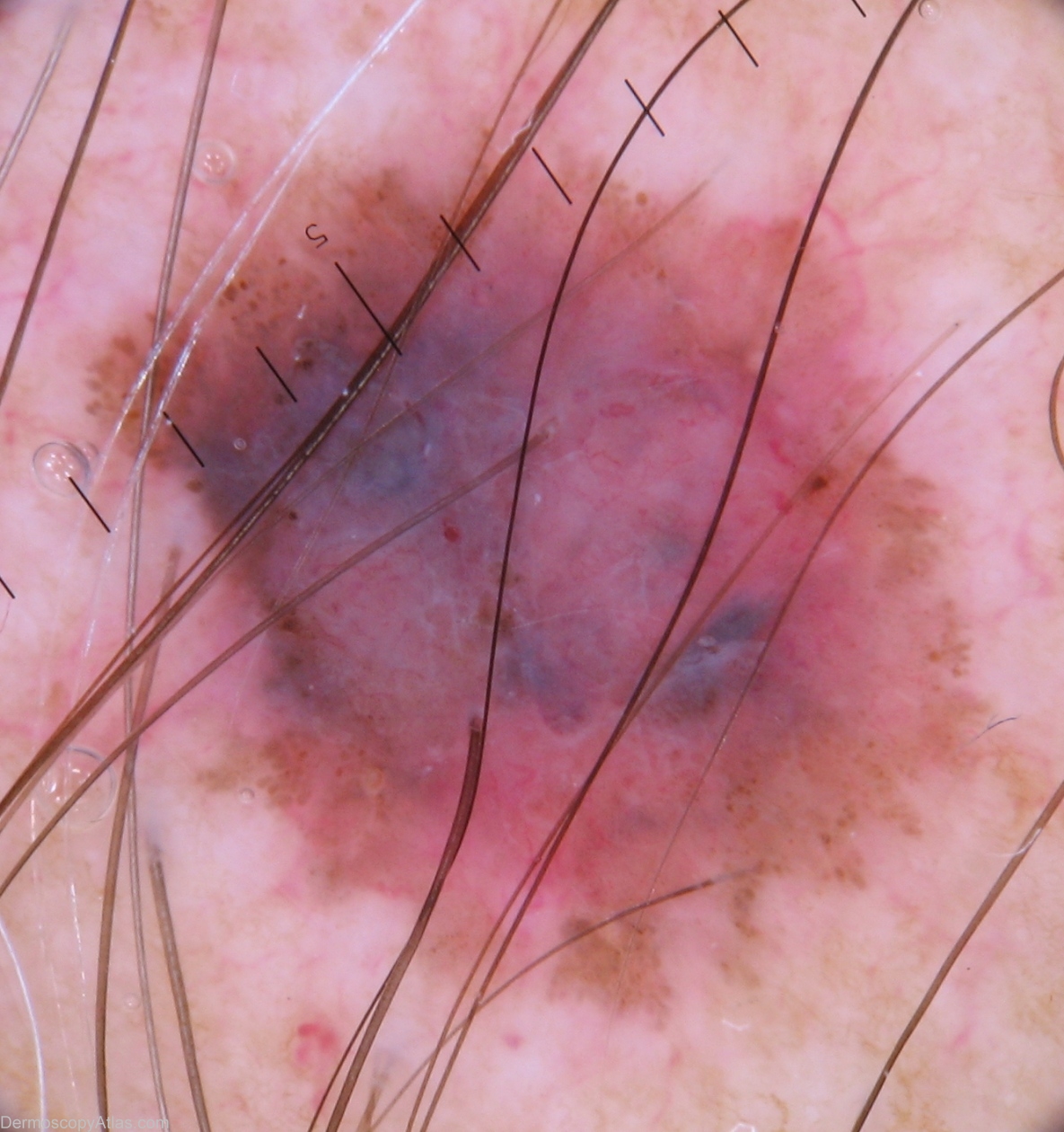
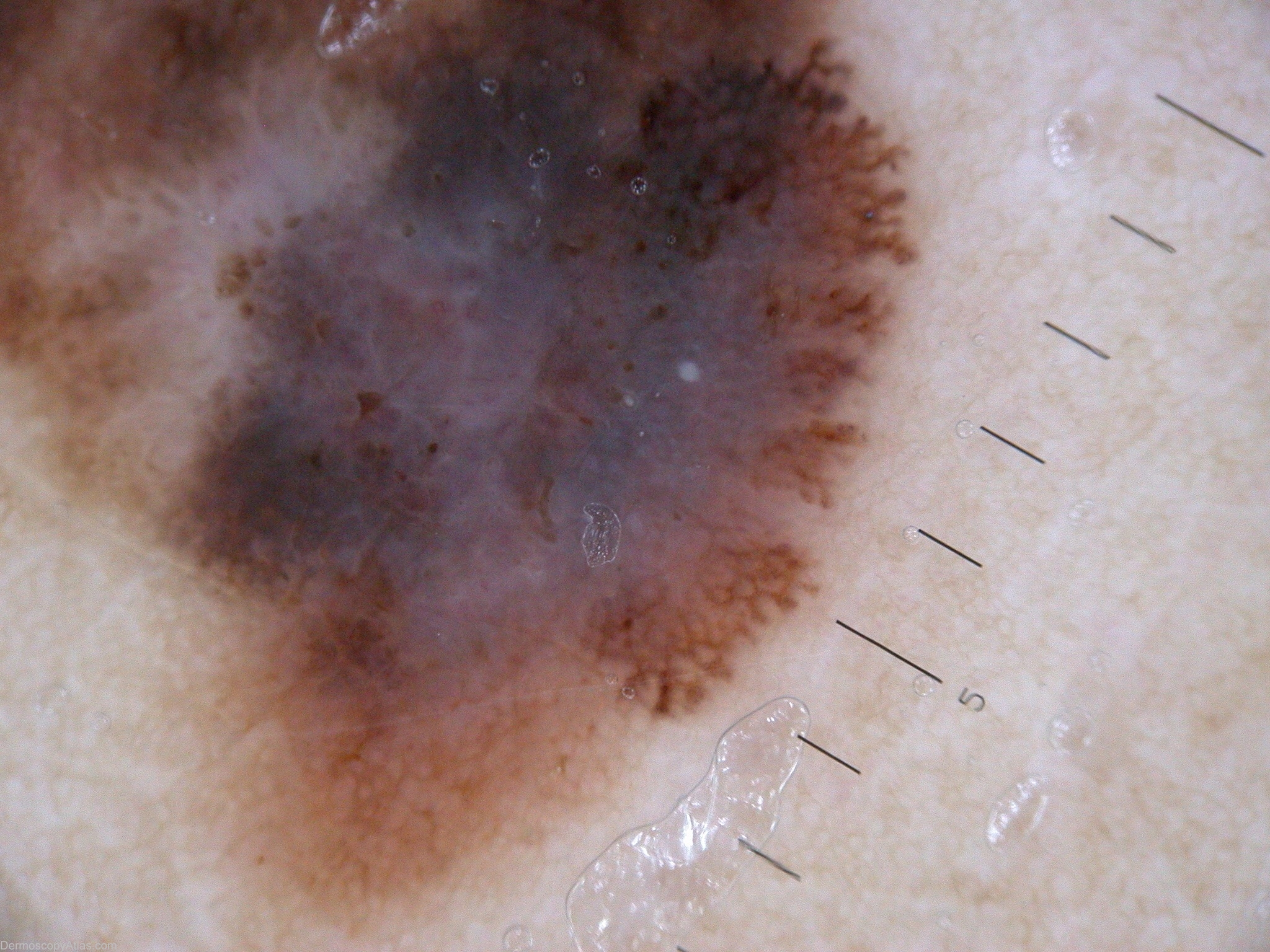
In comparison Melanoma shows
- Atypical Network: Lines are thickened and irregularly spaced.
- Blue-Whitish Veil: A hazy, structureless area over blue-gray pigmentation.
- Irregular Pseudopods/Streaks: Bulbous or finger-like projections at the edge.
- Atypical Vascular Pattern: A chaotic arrangement of vessels rather than clear branching.
See Dermoscopy Atlas for Pigmented Nodular Melanoma
See Dermoscopy Atlas for other Invasive melanomas
×
![]()